Treatment of retina and vitreous diseases
Retina is the innermost layer of eye ball. The image is formed by the optical apparatus of eye over the retina . It is very delicate part. Common diseases and there treatment facilities available at our centre are as follows:-
Treatment of comman retina and vitreous diseases
1. Age related macular degeneration : (ARMD)
This is degeneration of central part of retina due to age (more than 60 years). Patient has painless decrease of vision. Advanced cases have rupture of blood vessels in the lesion and require laser treatment or intravitreal injections of anti VEGF agents.
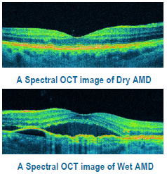
There are two types of AMD: dry (atrophic) and wet (neovascular or exudative). Most AMD starts as the dry type and in 10-20% of individuals, it progresses to the wet type.
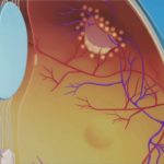
Dry ARMD;In dry age-related macular degeneration, small white or yellowish deposits, called drusen, form on the retina, beneath the macula, causing it to deteriorate or degenerate over time.
Wet ARMD;In wet age-related macular degeneration, abnormal blood vessels under the retina begin to grow toward the macula. Because these new blood vessels are abnormal, they tend to break, bleed, and leak fluid, damaging the macula and causing it to lift up and pull away from its base. This can result in a rapid and severe loss of central vision.
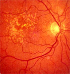
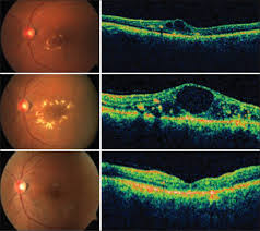
Dry ARMD
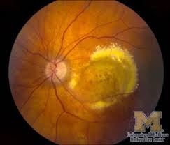
wet ARMD
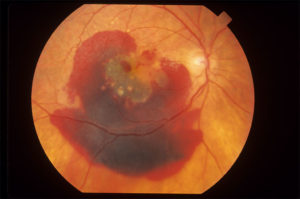
wet ARMD advanced
2. Diabetic retinopathy :
In patients of diabetes the retinal blood vessels become weak and proliferate producing bleeding and leakage. This produces decrease of vision or total loss of vision ( due to bleeding). Treatment is by intravitreal injections of anti VEGF agents,early laser [laser photo coagulation] or in some advanced cases vitreo retinal surgery.
The retina is the membrane that covers the back of the eye. It is highly sensitive to light.
It converts any light that hits the eye into signals that can be interpreted by the brain. This process produces visual images, and it is how sight functions in the human eye.
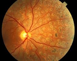
Diabetic retinopathy damages the blood vessels within the retinal tissue, causing them to leak fluid and distort vision.
There are two types of diabetic retinopathy DR:
- Non-proliferative diabetic retinopathy (NPDR): This is the milder form of diabetic retinopathy and is usually symptomless.
- Proliferative diabetic retinopathy (PDR): PDR is the most advanced stage of diabetic retinopathy and refers to the formation of new, abnormal blood vessels in the retina.

Diabetic retinopathy
At our centre latest equipment for treatment of diabetic retinopathy is available. After fundus fluorescein angioscopy the leaking blood vessels are sealed with green laser ( frequency doubled yag laser )For doing laser green laser is used.we are equipped with japneese latest nidek GYC1000 laser machine.


Green laser nidek GYC 1000

Laser photocoagulation
3.Anti-VEGF intravitreal injections
Your ophthalmologist may treat your wet ARMD,diabetic leakage in retina or other disease of the retina with injection inside the eye ,of a drug called anti-VEGF. Anti-VEGF treatment improves vision in about one third (1 out of 3) people who take it. For a vast majority (9 out of 10), it at least stabilizes vision.
What is Vascular Endothelial Growth Factor? VEGF
Vascular endothelial growth factor (VEGF) is a protein produced by cells in your body. VEGF produces new blood vessels when your body needs them.
Why Do You Want to Stop VEGF From Working?
Sometimes cells can produce too much VEGF. When this happens, abnormal blood vessels can grow in your eye. These abnormal blood vessels damage your eye and harm your vision. This can lead to low vision or blindness.
What Conditions Are Treated with Anti-VEGF Medicine?
Anti-VEGF medicine blocks VEGF, slowing the growth of blood vessels in the eye. This slows or stops damage from the abnormal blood vessels and slows down vision loss. Sometimes it can even improve vision.
Ophthalmologists use anti-VEGF medicines to treat the following eye problems:
- Wet age-related macular degeneration (AMD)
- Swelling of the retina, called macular edema
- Diabetic retinopathy
- Retinal vein occlusion
comman anti-VEGF medicines are
- Avastin
- Lucentis
4.Retinal blood vessel blockage diseases.
What is retinal vascular blockage?
Retinal blood vessel occlusion affects the eye, specifically the retina. The retina is the light-sensitive layer of tissue that lines the back of your eye. It’s covered with special cells called rods and cones that convert light into neural signals and send these signals on to the brain so you can see. The retina is vital for vision.
The vascular system includes blood vessels called arteries and veins, which transport blood throughout your body, including your eyes. Your retina requires a constant supply of blood to make sure your cells get enough nutrients and oxygen. Blood also removes the waste your retina produces. However, it’s possible for one of the vessels carrying blood to or from the retina to become blocked or to have a blood clot. This is called an occlusion.
The occlusion can cause blood or other fluids to build up and prevent the retina from properly filtering light. When light is blocked or fluids are present, a sudden loss of vision can occur. The severity of vision loss may depend on where the blockage or clot occurred.
Retinal vascular occlusion is a potentially serious condition, especially if hardening of the arteries, or atherosclerosis, already exists. It most often occurs in middle-aged and older people.
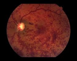
CRVO
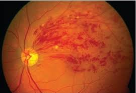
BRVO
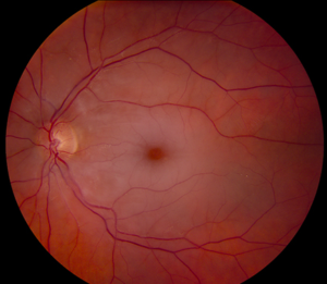
CRAO
What are the different types of retinal vascular occlusion? [see photos above]
There are two types of retinal vascular occlusion. The type depends on which blood vessel is affected:
1.Retinal vein occlusion
Retinal vein occlusion is blockage of one of your retinal veins, which are blood vessels that carry deoxygenated blood back to your heart. Retinal vein occlusion is also divided into two types:
- Central retinal vein occlusion (CRVO) is a blockage in the main vein of your retina, which is called the central retinal vein.
- Branch retinal vein occlusion (BRVO) occurs when the blockage is in a smaller branch of veins throughout the retina.
2.Retinal artery occlusion CRAO
Retinal artery occlusion is a blockage of one of the retinal arteries, which are blood vessels that carry oxygenated blood from the heart to your retina. A blockage in the main artery of your retina is called a central retinal artery occlusion. A branch retinal artery occlusion happens when the blockage occurs further along in the smaller branches of your artery.
Blockages in your main vein or artery are often more serious than blockages in your branch veins or arteries.
commonest causes are blood pressure,diabetes,high cholesterol,blood clots
Treatment; anti VEGF injections in eye,laser photocoagulation[ varies from case to case]
5.Retinal Detachment
If the retina gets separated from underlying layers, the condition is known as retinal detachment. the patient complains of flashes of light in front of eye, floaters and sudden decrease of vision or sensation of curtain coming from one side. Treatment is by surgery. In fresh and uncomplicated cases conventional retinal detachment surgery suffices. But in complicated and advanced cases vitreoretinal surgery and silicon oil injection etc is required.

Retinal Detachment

Retinal Detachment Surgery by putting silicon buckle

Vitroretinal Surgery
Prophylaxis of retinal detachment : If breaks in the retina are detected before retina has actually detached, these breaks can be sealed with laser or cryotherapy (very cold temperature)
Laser sealing of breaks

Cryotherapy
6. Sunken Cataractous Lens or Intraocular Lens (IOL) or foreign body:
Some times due to trauma the lens of eye falls back into cavity of eye, similarly during surgery cataractous lens or intraocutor lens may fall back. In such cases the jelly inside the eye (vitreous) is removed (vitrectomy) a special liquid is put inside the eye ball (PFCL) so that cataractous lens or IOL which has fallen back comes to float over it and it is removed.
Due to trauma foreign bodies may go inside the eye ,these can also be removed by VR surgery
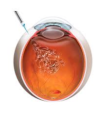
6. Vitreous Hemorrhage :
Bleeding into the jelly filling the cavity of eye (Vitreous) is known as vitreous haemorrhage. Common causes of vitreous haemorrhage are diabetic retinopathy and trauma. Vitreous haemorrhage may get reabsorbed in few months. If it does not get reabsorbed vitrectomy surgery is done to remove the vitreous and blood.

Vitrectomy
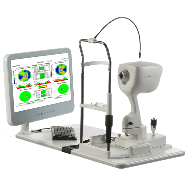
OCT..the diagnostic tool for diseases of retina
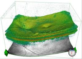
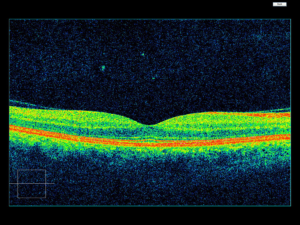
Optical coherence tomography (OCT) is a non-invasive imaging test. OCT uses light waves to take cross-section pictures of your retina.
With OCT, your ophthalmologist can see each of the retina’s distinctive layers. This allows your ophthalmologist to map and measure their thickness. These measurements help with diagnosis. They also provide treatment guidance for glaucoma and diseases of the retina. These retinal diseases include age-related macular degeneration (AMD) and diabetic eye disease,vascular occlusions as discussed above.
What happens during OCT?
To prepare you for an OCT exam, your ophthalmologist may or may not put dilating eye drops in your eyes. These drops widen your pupil and make it easier to examine the retina.
You will sit in front of the OCT machine and rest your head on a support to keep it motionless. The equipment will then scan your eye without touching it. Scanning takes about 5 – 10 minutes. If your eyes were dilated, they may be sensitive to light for several hours after the exam.

I am text block. Click edit button to change this text. Lorem ipsum dolor sit amet, consectetur adipiscing elit. Ut elit tellus, luctus nec ullamcorper mattis, pulvinar dapibus leo.